To the untrained eye, a cryo-electron tomogram seems to be traces in sand rather than a comprehensive photograph of a cell.
These pictures may be used by specialists trained in sophisticated microscopy methods like as cryo-electron microscopy and tomography to analyse the location and form of cellular organelles as well as the structures of massive molecular complexes. As a consequence, scientists may get insight into the inner workings of a cell in both healthy and pathological states.
However, there is a significant disadvantage to this strategy. While qualified professionals are capable of distinguishing and classifying various cellular formations in tomograms, the technique is exceedingly time-consuming.
This is why the EMBL Heidelberg groups of Zaugg, Mahamid, Kreshuk, and Diz-Muoz developed an artificial intelligence-based system for fast and effectively annotating cellular structures in cryo-electron tomograms. They presented this tool in a recent paper in Nature Methods, and it is now freely accessible and used by the scientific community.
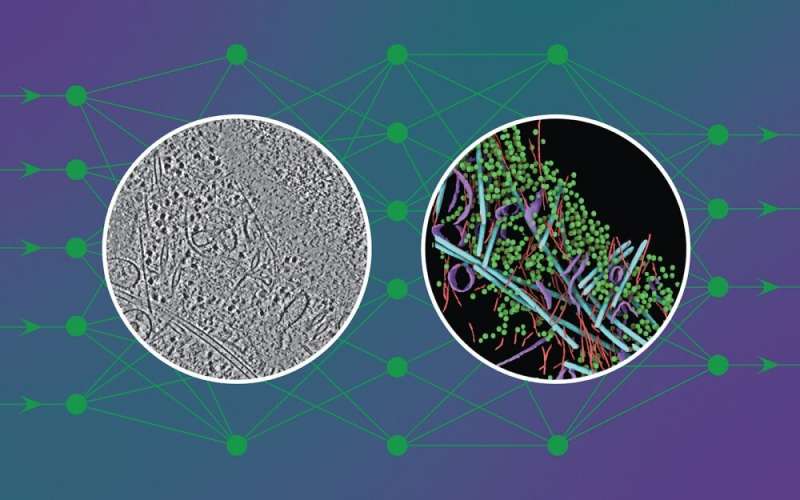
Deep-learning framework DeePiCt (Deep Picker in Context) can distinguish and name organelles and chemical complexes far quicker than the human eye and without human bias, creating highly detailed cellular pictures (like the one in the circle on the right in the image above).
“DeePiCt—specifically, the trained models that we provide—allows anybody to discover certain particles and structures of interest within the noisy background of their own tomograms. This is one of the greatest outputs of our effort in my opinion “Judith Zaugg said. “Without it, you had to call a skilled professional for assistance with annotations, which may take a long time. DeePiCt, in my opinion, is a crucial step toward allowing high-throughput in cell structural biology.”
The DeePiCt framework enables scientists to readily categorise cellular features in tomograms depending on their location inside the cell. This may then be used to compare the class of ribosomes found on mitochondria to those found on the endoplasmic reticulum, for example. Such studies have already revealed previously unknown structural information about how ribosomes bind to these various membranes.
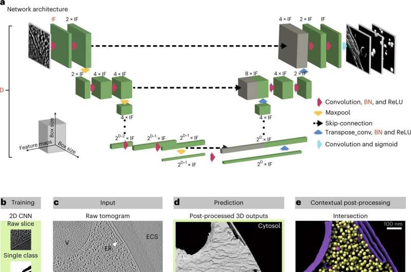
Two kinds of convolutional neural networks are used in the programme. These are deep-learning algorithms that can detect patterns and distinguish items in images. The first, which functions in 2D slices, was taught to segment cellular features such as organelles and cytoplasm. The second, which works in the three-dimensional space of a tomogram, was taught to segment a particle of interest (e.g., a ribosome).
Importantly, once taught to detect a given particle in a set of tomograms, the network was able to identify the same particles in fresh tomograms, even those of cells from a different species. This implies that DeePiCt can be utilised by cryo-electron tomography researchers on a wide range of sample types.
The network was trained to recognise four unique structures (actin, ribosomes, microtubules, and membranes) in cells from three different creatures in order to predict these features in an unseen tomogram from a human cell in the figure displayed.
“Now that we’ve shown that this works, we’re pleased to make the programme open to the scientific community,” Julia Mahamid said. “We expect that deep-learning methods like this will become the gold standard in cryo-electron tomography. In addition, we are putting 20 well-annotated tomograms in the EMBL-EBI archives, which we hope will spark and promote future technique development in the scientific community.”
You might also be interested in reading, Researchers develop a novel way of creating spinning thermal radiation