Researchers at the University of Alabama in Birmingham used cryo-electron microscopy to reveal the structure of a bacterial virus in unprecedented detail. This is the first structure of a virus capable of infecting Staphylococcus epidermidis, and high-resolution structural information provides a critical connection between viral biology and the virus’s potential therapeutic usage to treat bacterial diseases.
Bacteriophages, sometimes known as “phages,” are viruses that infect bacteria. Terje Dokland, Ph.D., of the University of Alabama at Birmingham, and Asma Hatoum-Aslan, Ph.D., of the University of Illinois Urbana-Champaign, have developed atomic models for all or part of 11 distinct structural proteins in phage Andhra. The findings were reported in Science Advances.
Andhra belongs to the picovirus family. It only has S. epidermidis as a host. This skin bacteria is typically harmless, although it is a significant source of indwelling medical device infections. “Picoviruses are infrequently identified in phage collections and are understudied and underutilised for therapeutic applications,” Hatoum-Aslan, a phage scientist at the University of Illinois, stated.
With the advent of antibiotic resistance in S. epidermidis and the related pathogen Staphylococcus aureus, scientists have rekindled their interest in employing bacteriophages to treat bacterial infections. Picoviruses invariably destroy the cells they infect after attaching to the bacterial cell wall, breaking through it enzymatically, piercing the cell membrane, and injecting viral DNA into the cell. They also have additional characteristics that make them appealing therapeutic possibilities, such as a tiny genome and the difficulty to transmit bacterial genes across bacteria.
Understanding protein structure in Andhra and how those features help the virus to infect a bacteria would allow genetic modification to create custom-made phages customised to a certain purpose.
“The structural basis for host specificity between phages that infect S. aureus and S. epidermidis remains unknown,” said Dokland, a UAB professor of microbiology and head of the UAB Cryo-Electron Microscopy Core. “The current work has improved our knowledge of the structures and activities of Andhra gene products, as well as the determinants of host specificity, opening the path for more rational creation of bespoke phages for therapeutic purposes. Our results provide light on essential aspects of virion assembly, host identification, and penetration.”
Staphylococcal phages generally infect a limited number of bacteria due to the varied polymers of wall teichoic acid on the surface of various bacterial strains. “On the one hand, this restricted host range permits the phages to target just the individual pathogen causing the sickness; on the other hand, it implies that the phage may need to be customised to the patient in each unique instance,” Dokland said.
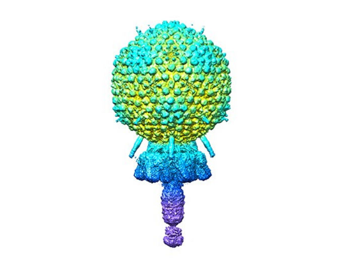
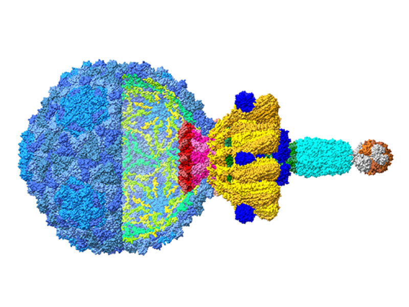
Andhra’s general structure is a 20-faced, roundish icosahedral capsid head containing the viral DNA. A short tail is joined to the capsid. The tail is mostly responsible for attaching to S. epidermidis and enzymatically shattering the cell wall. The viral DNA is introduced into the bacteria through the tail. Segments of the tail include the portal from the capsid to the tail, and the stem, appendages, knob and tail tip.
Each viral particle’s 11 distinct proteins are present in many copies that assemble together. The capsid, for example, is made up of 235 copies of two proteins, while the remaining nine virion proteins have copy counts ranging from two to 72. The virion is composed of 645 protein components, including two copies of a 12th protein, the structure of which was predicted using the protein structure prediction tool AlphaFold.
The atomic models described by Dokland, Hatoum-Aslan, and co-first authors N’Toia C. Hawkins, Ph.D., and James L. Kizziah, Ph.D., both of the University of Alabama at Birmingham Department of Microbiology, show the structures for each protein, as described in molecular language such as alpha-helix, beta-strand, beta-barrel, or beta-prism. The researchers have defined how each protein links to other copies of the same protein type, for example, to form the capsid’s hexameric and pentameric faces, as well as how each protein interacts with neighbouring distinct protein types.
Electron microscopes illuminate an item with a stream of accelerated electrons, offering significantly better resolution than a light microscope. Cryo-electron microscopy adds the aspect of super-cold temperatures, making it especially effective for near-atomic structural resolution of bigger proteins, membrane proteins, or lipid-containing materials such as membrane-bound receptors, as well as complexes of multiple biomolecules.
Over the last eight years, new electron detectors have resulted in a significant increase in resolution for cryo-electron microscopy over conventional electron microscopy. The following are key components of the so-called “resolution revolution” in cryo-electron microscopy:
- Aqueous samples flash-frozen in liquid ethane chilled to less than -256 degrees F. Water freezes to a window-like “vitreous ice” instead of ice crystals, which damage samples and scatter the electron beam.
- To minimise protein damage, the material is held at super-cold temperatures in the microscope, and a low dosage of electrons is employed.
- Direct electron detectors that are very quick may count individual atoms at hundreds of frames per second, enabling sample movement to be adjusted on the fly.
- Advanced computing combines dozens of photos to create high-resolution three-dimensional structures. Terabytes of data are processed by graphics processing units.
- The microscope stage, which holds the sample, may also be tilted while pictures are captured, enabling the creation of a three-dimensional tomographic image, comparable to a CT scan at the hospital.
The UAB researchers began their examination of the Andhra virion structure with 230,714 particle pictures. The 186,542, 159,489, 159,489, and 159,489 pictures were used to begin the molecular reconstruction of the capsid, tail, distal tail, and tail tip, respectively. The resolution varied between 3.50 and 4.90 angstroms.