For high-resolution imaging of deep tissues, photoacoustic tomography (PAT), a hybrid imaging approach, combines optical illumination with ultrasonic detection. PAT offers a clear benefit with scaling resolution, greater imaging depth, and high contrast imaging by utilising the photoacoustic (PA) effect.
It illuminates the target tissue with nanosecond laser pulses so that the chromophores can take up the incident laser energy. This causes a localised increase in temperature and creates pressure waves that travel as ultrasonic waves to the tissue boundary. Using reconstruction methods, these PA waves are subsequently captured with the aid of an ultrasonic transducer and transformed into internal absorption maps.
The simple delay-and-sum (DAS) beamformer is one of many image reconstruction methods that can be used to generate the initial pressure maps. The recorded signals from various tissue sites are back-projected by this technique, and they are then added to each pixel in the reconstructed image.
However, this increases the computational cost and processing time of the DAS beamformer and causes artefacts, or anomalies, in the reconstructed pictures. Despite these shortcomings, it is a popular option for PAT reconstruction due to its simplicity and ease of application.
Implementing these reconstruction algorithms typically calls for a workstation, desktop, or laptop with powerful processing capabilities. However, in recent years, mobile phones’ processing capability has increased.
Although the use of mobile phones for other microscopy techniques, such as ultrasound imaging, has been suggested, its potential for photoacoustic imaging techniques like PAT image reconstruction has not been investigated.
Researchers from Singapore and the United States have now created an Android-based application for PAT picture reconstruction, capitalising on the powerful computational power of mobile devices.
Manojit Pramanik, a Northrop Grumman Associate Professor in Iowa State University’s Department of Electrical and Computer Engineering, directed the work, which was written up in the Journal of Biomedical Optics (JBO).
On Kivy, a cross-platform Python 3.9.5 framework, the created application uses a single-element ultrasonic transducer (SUT)-based DAS beamformer technique for image reconstruction.
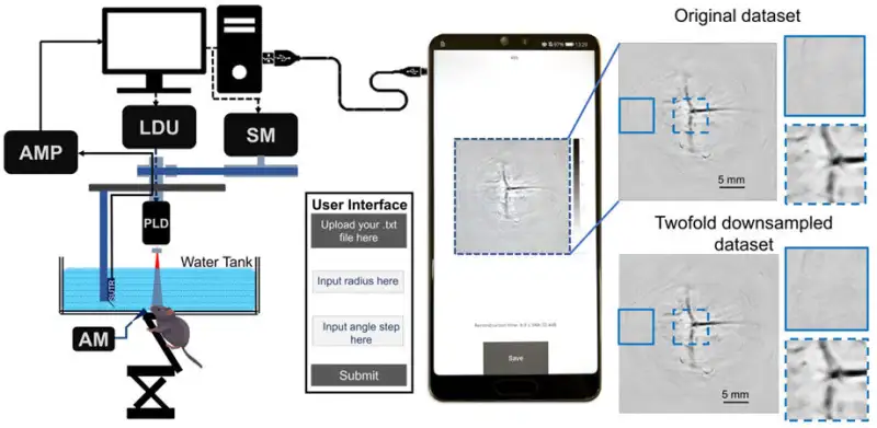
Using simulated and experimental PAT datasets, the researchers evaluated its performance on various mobile phones. The experimental datasets included a point source phantom, a triangular form phantom, and blood vessels in the brains of live rats, whereas the simulation datasets included point targets, triangular shapes, and rat’s brain vessel shapes.
“The developed application can successfully reconstruct the PAT data into high-quality PAT images with signal-to-noise ratio values above 30 decibels,” comments Pramanik.
It’s interesting to note that for small datasets, the processing time of the algorithm on a Huawei P20 mobile phone was comparable to that on a laptop.
Additionally, two-fold downsampling of the original dataset decreased the computational time while keeping the image quality, enabling for fast and high-quality image reconstruction. In comparison, the PAT photos were noticeably worse after three-fold downsampling.
Furthermore, the researchers discovered that PAT reconstruction could be completed in just 2.4 seconds because to the Samsung Galaxy S21+’s cutting-edge chipset. The efficiency of the mobile phone application is shown by the significantly shortened running time for image reconstruction, according to Pramanik.
JBO Editor-in-Chief Brian Pogue, Chair of Medical Physics at University of Wisconsin–Madison, remarks, “This first-of-its-kind application provides an opportunity for PAT image reconstruction on inexpensive, portable, and widely available mobile phones.
Going ahead, the application can make PAT systems more adaptable and extendable to other fields of biomedical imaging, facilitating point-of-care diagnosis.” He adds, “The code for this Android-based application has been made freely available on GitHub, making this a major service to the biomedical imaging community.”